“mahala” the Florida Panther
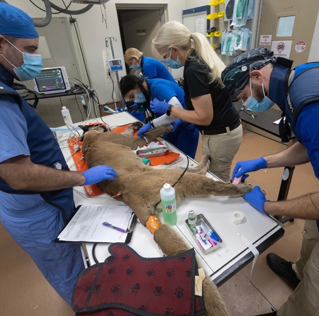
With a great start to my first week at Zoo Miami, the 10-year-old Florida Panther “Mahala” underwent a preventative health exam, including an ovariohysterectomy.
As intact felines grow older they become susceptible to complications such as pyometra (a uterine infection) and various forms of uterine cancer.
Since Mahala is a federally protected, non-releasable animal & not apart of the breeding program, spaying her is the best way to prevent any future complications in her reproductive tract.
In addition to the ovariohysterectomy, Mahala also received bloodwork, radiograph imaging, and a dental cleaning. For the safety of zoo staff and animals, it is best to perform immobilizations & preventative health exams only when absolutely necessary.
Preparing for Examinations
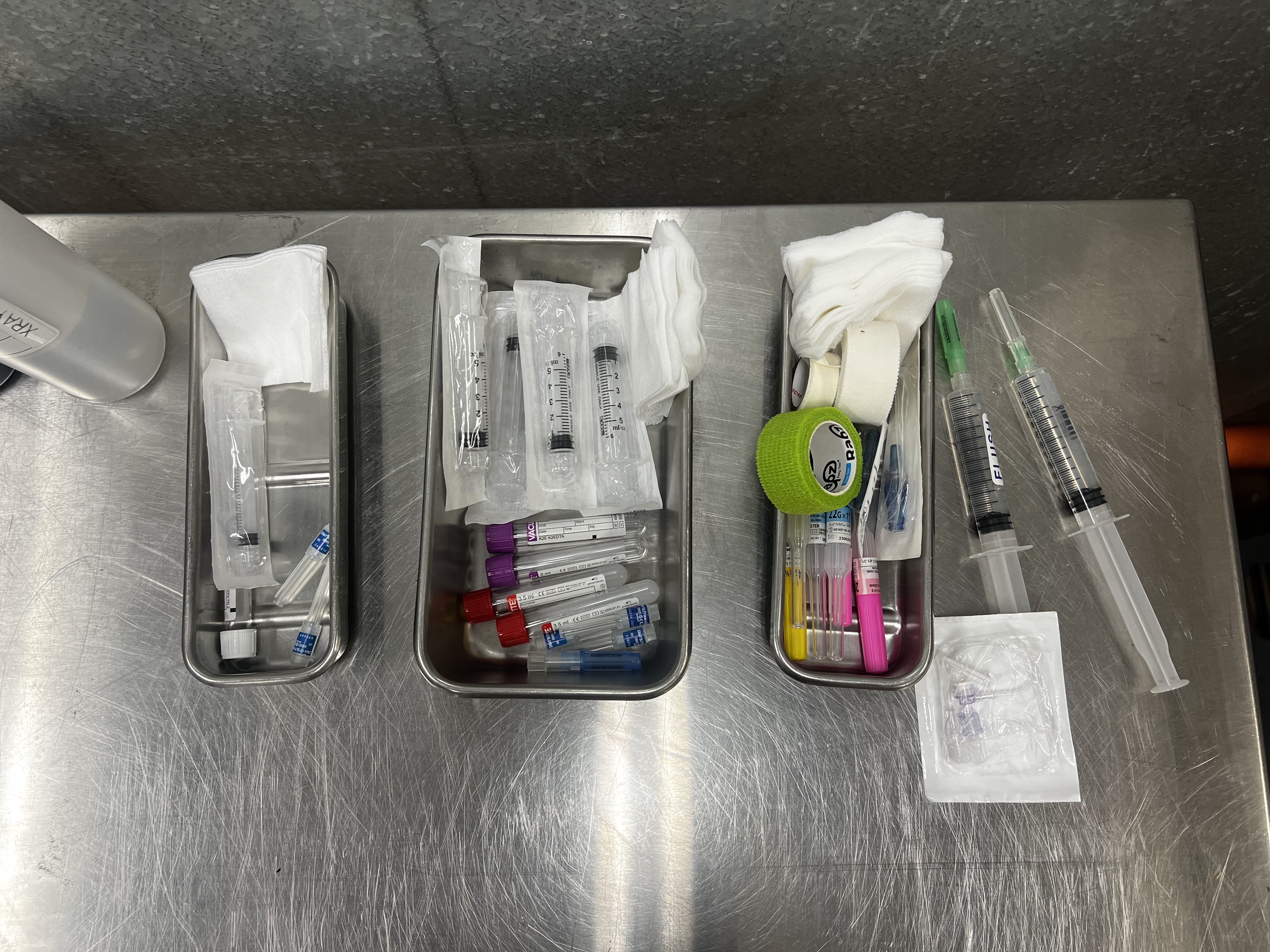
One of my first duties as a clinical intern was to prepare the necessary items for routine exams. Most often, standard pre-shipment and annual exams of the zoo collection animals consisted of catheter placement, blood collection, and in some instances, urine collection.
After reading through the examination plan on the daily schedule, I would begin to assist the veterinary technicians in preparing the things needed. On the left are the supplies needed for a cystocentesis, or a sterile urine collection that is taken directly from the bladder with a needle. The things placed in the middle consist of blood collection tubes (EDTA & Clot Activator), syringes, and needles to collect blood from the animal that will be biochemically analyzed. The size of the syringes used depends on the size of animal and the amount of blood being collected. The tools on the right are going to be used to place a catheter into the animal. The catheters themselves are the yellow and pink colored caps (the color varying with the size of the needle). You will also need materials such as gauze, tape, & Vetwrap to hold the catheter in place once it is inserted intravenously. The blue colored cap is used to plug the base of the needle once blood enters and the T-port extension set is used to aid in blood collection and the delivery of fluids and other medications through the catheter. LRS fluid is also prepared, kept warm, and labeled as”flush” to flush the catheter clean.
Blood Collection Tubes

Serum
A red top can either be a plain tube or contain a clot activator to yield serum. When blood clots it becomes separated into about 50% clotted red blood cells and 50% plasma. Plasma – clot = serum which is achieved by spinning the sample down in a centrifuge for about 15 minutes. The spun blood separates the serum from the clot. Serum biochemistries are important because this liquid contains proteins, enzymes, & lipids which can be useful in determining the presence of disease.
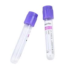
K2 EDTA
The walls of a purple top tube are lined with EDTA, an anticoagulant. EDTA binds calcium ions which prevents the blood from clotting. This causes the blood to remain in a liquid state which preserves red and white blood cell morphology. This is useful for tests that analyze the number of cells like CBCs. It can also be used to examine blood cell morphology or blood typing. Both red/tiger or purple/lavender tops are used in mammal blood analysis.

Lithium Heparin
In green tops, heparin is used as an anticoagulant by enhancing antithrombin III which inhibits thrombin and other clotting factors. This allows the blood to stay in a liquid state which is useful for tests examining whole blood or plasma. These tubes are also commonly used for chemistry analysis of factors such as arterial blood gases and plasma electrolytes. If wanting to do a basic blood analysis, such as a CBC or biochemistry in a reptile or a bird, green heparin tubes are most commonly used. This is because their blood is nucleated and EDTA can disrupt the cell morphology. Their blood also clots inconsistently, so it is more reliable to use an anti-coagulant like heparin to analyze plasma.
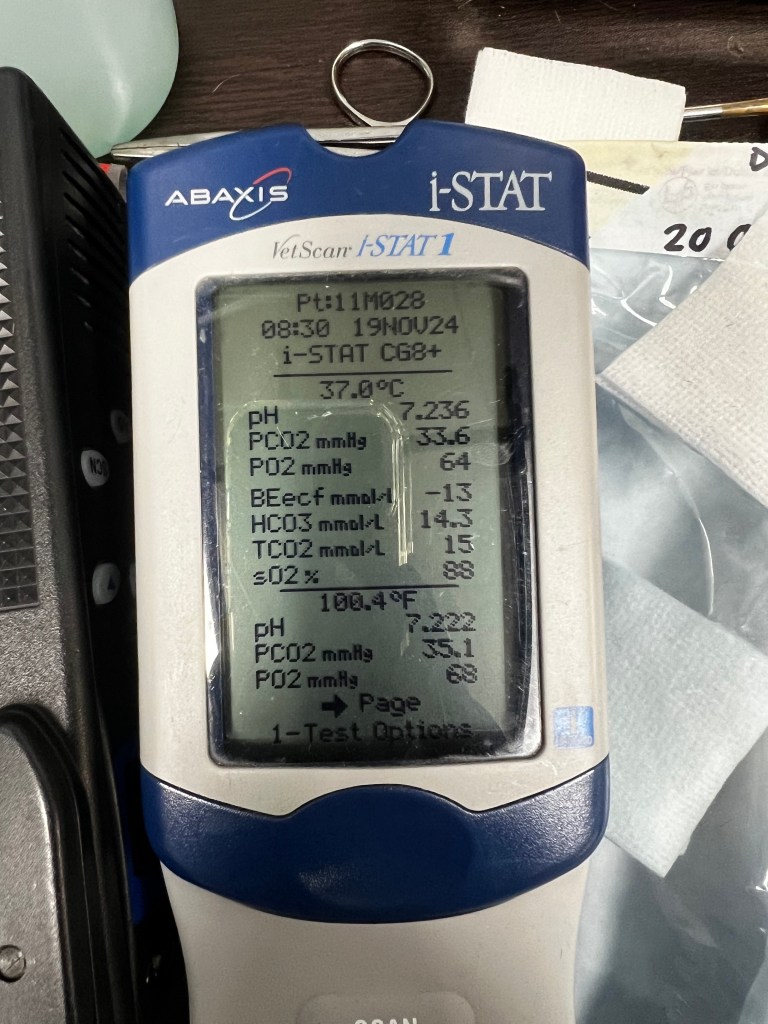
i-STAT
The i-STAT is a machine used to analyze various diagnostics such as blood gases, chemistries/electrolytes, lactate levels & more, of both mammalian and reptilian patients in real-time. With the use of an i-STAT cartridge and as little as 2-3 drops of whole blood, results can be produced within 2 minutes! It’s pretty cool to be able to see a patient’s blood analysis while they’re still on the table.
Common Anesthetics & Reversals
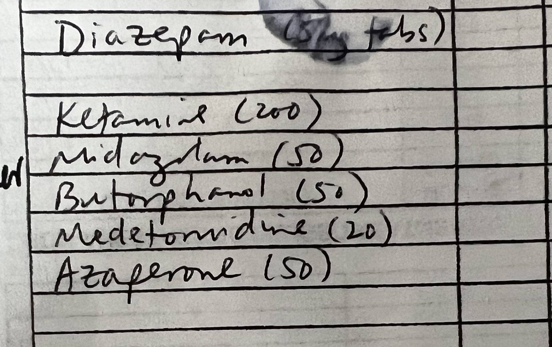
Anesthetics
In veterinary medicine, as in human medicine, it is crucial to document the exact time, dosage, and volume of sedatives administered to the patient. Sedation is very commonly used in zoo/exotic wildlife procedures and exams due to how stressful & dangerous it can be on the animals and zoo team to handle them. Of course the anesthetic drugs used vary depending on the size and species of the patient as well. Here are some common drug mixtures used to sedate large mammals.
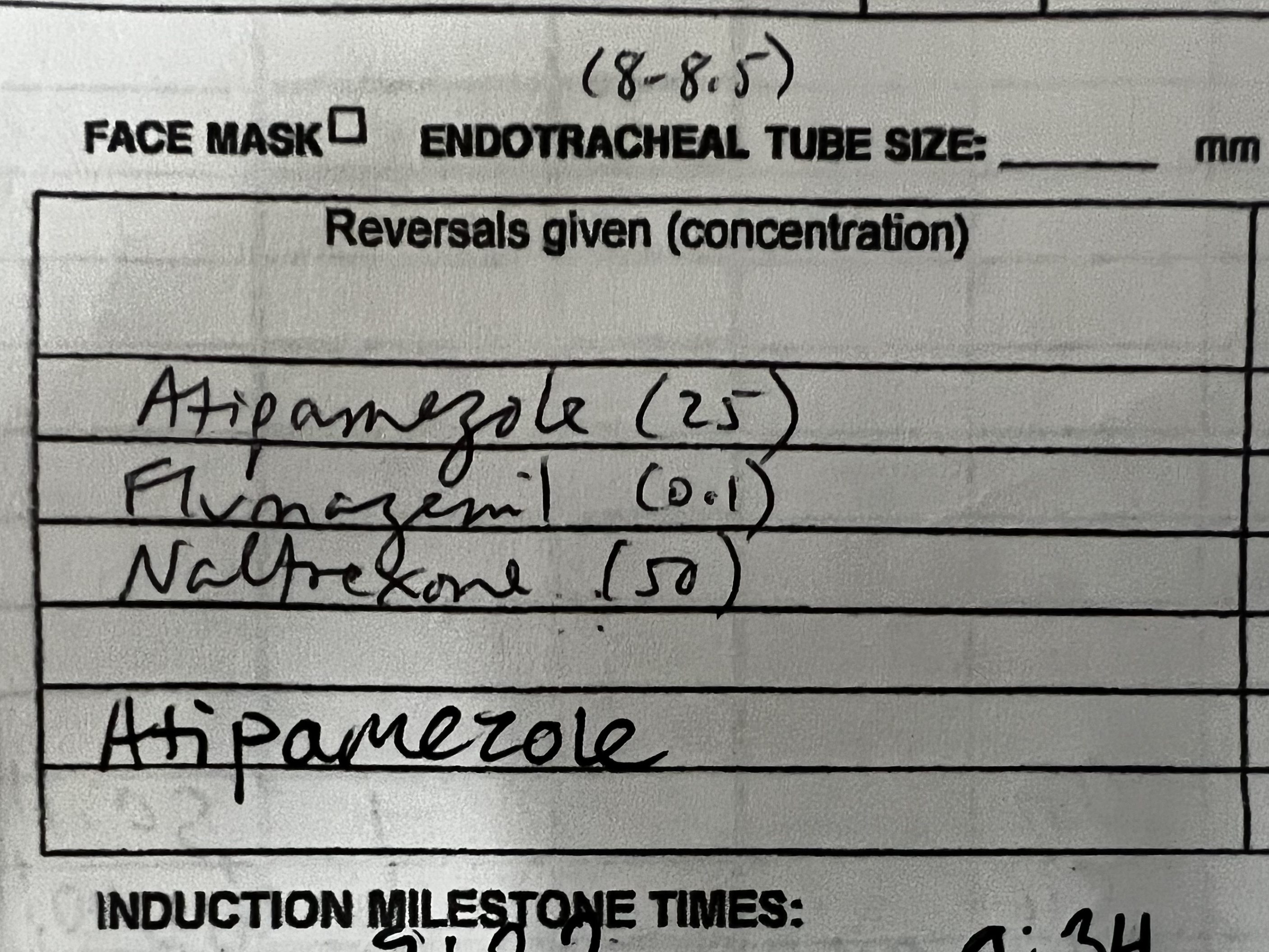
Reversals
Most drugs used for sedation also have an associated “reversal” compound that is used to wake the patient up after a procedure. Atipamezole, also commonly known as Antisedan, is a reversal agent of alpha2-adrenergic agonist drugs such as Medetomidine. Flumazenil is another commonly used reversal agent for benzodiazepines, which in this case are the Midazolam & Diazepam. The third reversal present in this image, Naltrexone, is a rapid reversal agent of opioids such as Butorphanol.
Some Unique Anatomy!
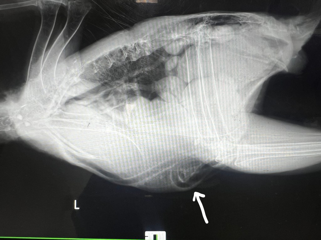
Some bird species, like cranes, swans, and guineafowl possess a unique anatomical trait referred to as “tracheal elongation” or tracheal looping. This occurs when the trachea is longer than typical and oftens loops within the chest cavity which can be seen in this radiograph of a guineafowl! It is thought that this anatomical feature aids in acoustic signaling and communication.
Ulcerative Pododermatitis
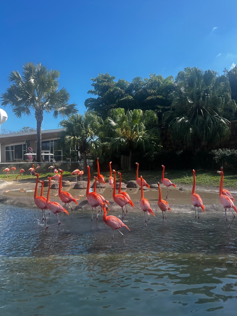
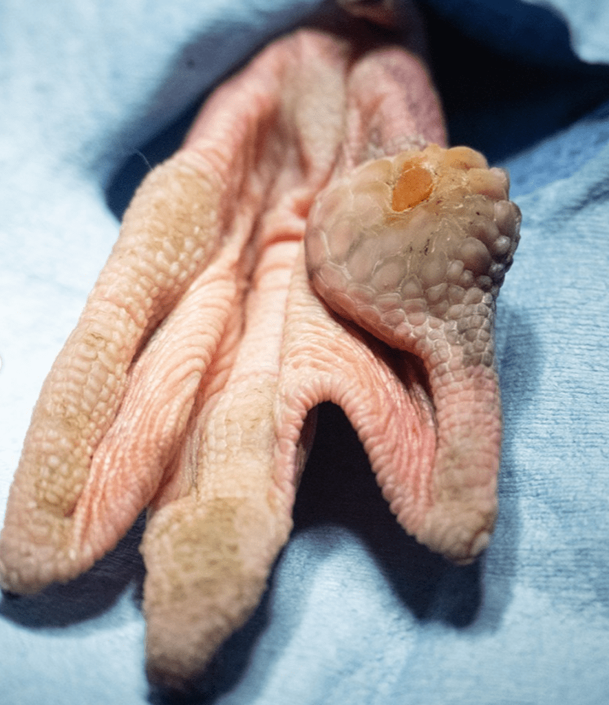
From my time at Zoo Miami, I learned that many avian species are susceptible to a bacterial infection called ulcerative pododermatitis, or bumblefoot. It involves either one or both of the footpads and is commonly found in birds like chickens, birds of prey, and apparently flamingoes. This condition begins to appear as a firm, inflammatory lesion on the bottom of the foot. It typically stems from a wound or irritation from an abrasive surface, wet bedding, or overgrown toenails which then turns infected. These regions may eventually become ulcerated if left untreated and lead to the development of abscesses that can cause the bird to exhibit lameness and other systemic complications. This condition can require an extensive treatment plan depending on which stage it has developed.
Six of the flamingoes at Zoo Miami were found to have bumblefoot and were required to remain in the hospital’s ICU and receive veterinary treatment. In severe cases the masses need to be surgically removed, but in early stages it can typically be treated with foot wraps, oral anti-inflammatory, and antibiotic medication.
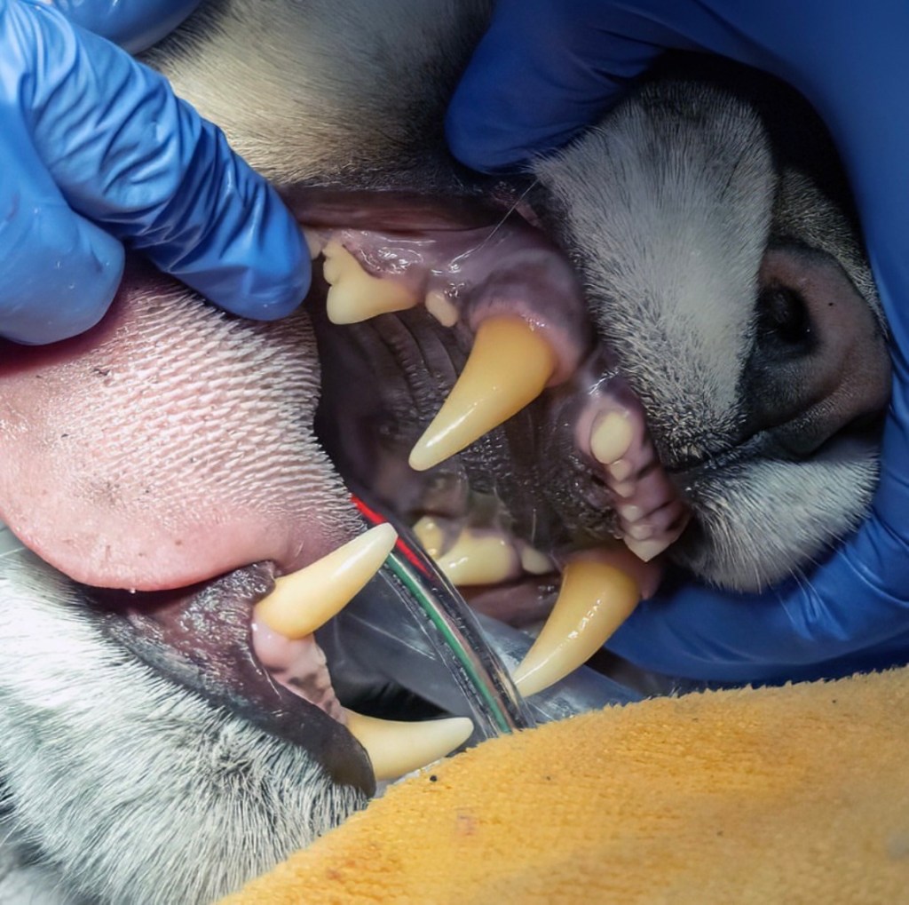
Dental Week! 🦷🪥
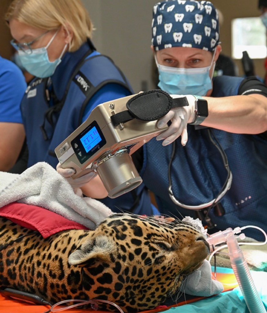
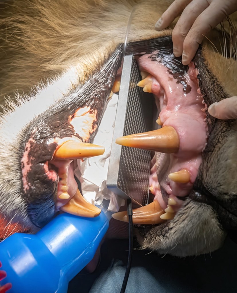

Just like us, animals need to receive routine veterinary dental care. Dr. Jamie Berning DVM, DAVDC is a board-certified veterinary dentist that will fly to Zoo Miami, often annually, to facilitate dental treatments and preventative care for various species of animals.
Zoo Miami’s dental week consists of dental procedures and treatment plans for at least five different animals. I was so lucky to have completed my internship and assist during the zoo’s 2025 dental week, which provided care for the following animals:
- Lion
- Orangutan
- Sloth bear
- North American River Otter
- Chimpanzees
- Jaguars
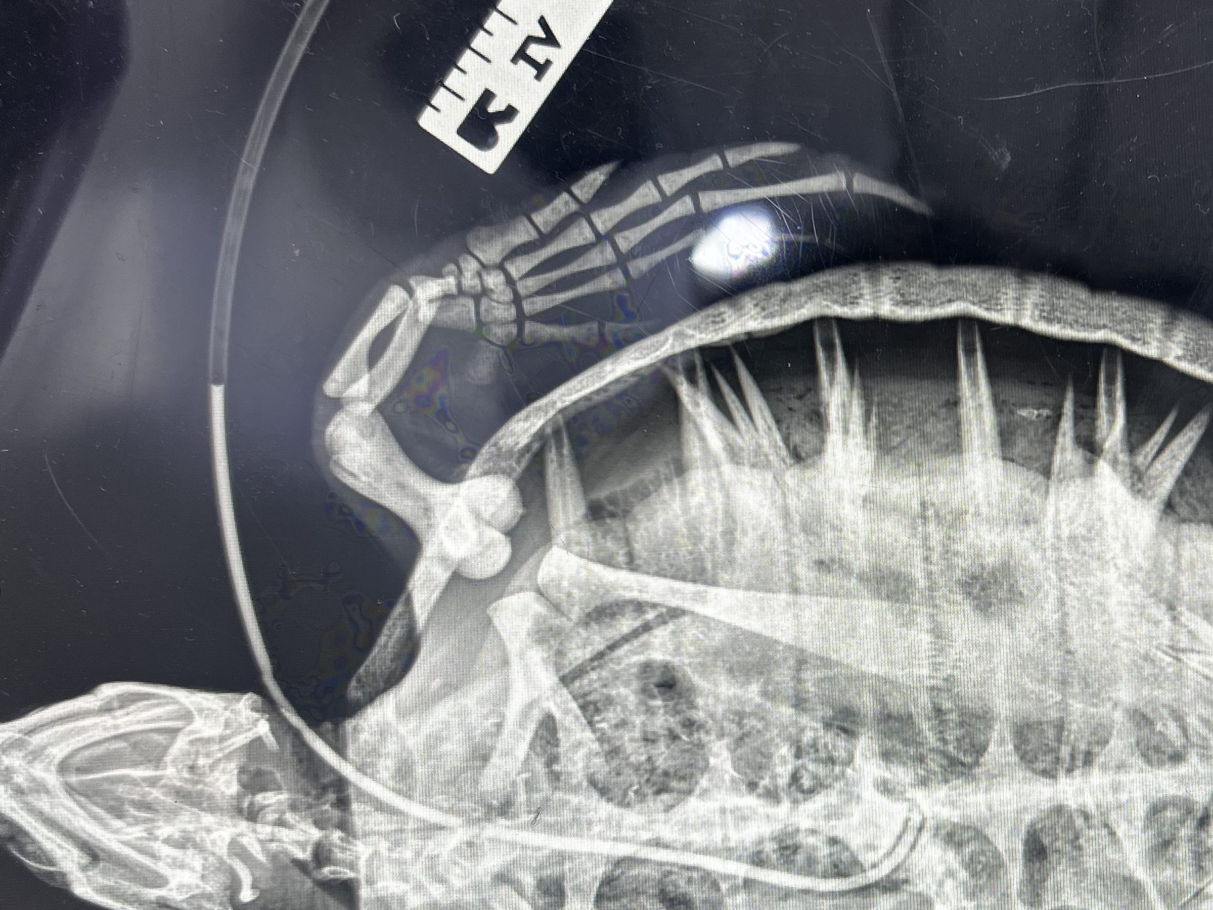
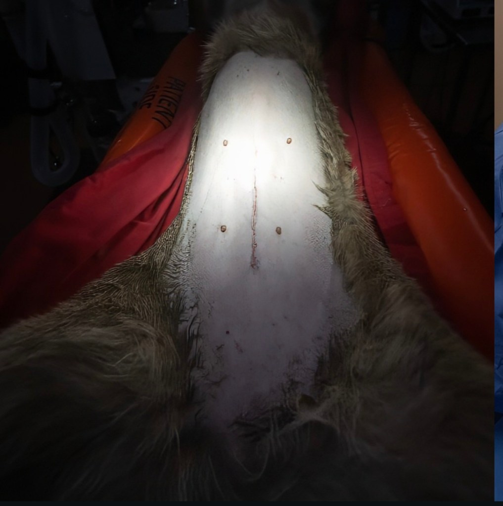
Proper dental assessments require radiographs, as pictured above, and for the patient to be sedated (especially when it’s a lion). A majority of the animals are trained to voluntarily receive the sedation via injection and are then transported up to the hospital with the help of the veterinary, keeper, and trained animal response teams.
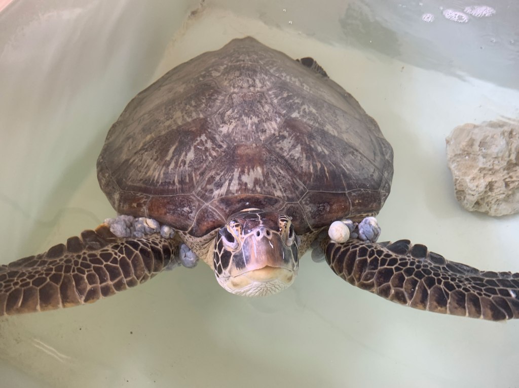
Zoo Miami’s Sea Turtle Hospital
Although Zoo Miami does not keep sea turtles as part of their collection, the Sea Turtle Hospital was recently built to aid in conservation and rehabilitation efforts for Sea turtle species from various regions around Florida. The turtles are transported to Zoo Miami where they will stay, receive proper veterinary care, and then be released back into the wild once they are strong and recovered.
During the colder months, turtles in the northern regions of Florida tend to experience “cold-stunning”, a condition in which the turtles become weak and immunosuppressed from exposure to the cold temperatures. A lot of turtles are also susceptible to a viral infection called Fibropapillomatosis, or FP, which results in the growth of proliferative masses along the turtle which can inhibit the ability to properly eat, swim, and avoid predation. Regardless of the reason, suffering turtles are found by various organizations along Florida’s eastern-coast regions and are transported to Zoo Miami for recovery.
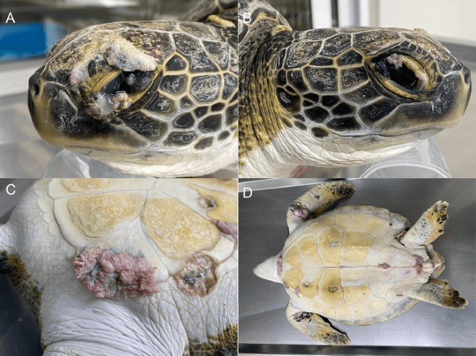
Fibropapillomatosis
FP is a neoplastic, infectious disease that has been reported to affect all seven species of sea turtles, but is most commonly reported in the green sea turtle. The etiological agent of FP is chelonid-herpesvirus 5 or ChHV5, a double-stranded DNA virus that is hypothesized to be transmitted through direct contact with contaminated surfaces, water, or other infected turtles. There is still a lot that is unknown about the origin, transmission, and progression of this disease but zoological vets all over the world work to treat and prevent it to the best of their ability.
Infection with ChHV5 has the potential to result in the growth of both internal and external tumors almost anywhere on the body. Although the tumors themselves are benign, it is the location in which they grow that leads to the animal’s debilitation. Tumors have the potential to grow in areas on the face, like the eyes, which can impact the turtle’s ability to forage for food and avoid predators. Large tumor growths around the mouth and extremities of the turtle can negatively impact it’s ability to swim and consume their diet leading to malnutrition. External growths are also commonly seen on the, cornea, carapace, and sutures between the scutes of the plastron. It is thought that subsequent to external growth, the tumors will begin to grow internally in areas like the oral cavity, esophagus, heart, lungs, GI tract, etc.
Depending on the severity of the growths, turtles can undergo surgical intervention to remove the impactful masses. There are various methods for tumor removal, including the use of a scalpel, electrocautery, and cryosurgery. It seems that to date, the standard approach to tumor removal involves the use of the CO2 laser. Although these images are not from Zoo Miami, I have included some examples of how the proliferative masses can grow and what they look like.
“Cold-stunning”
“Cold-stunning” is a condition experienced by sea turtles in which they become lethargic and weak due to exposure to cold water temperatures, typically below 50˚F (10˚C). Because sea turtles are ectotherms, they are unable to reach and maintain their adequate body temperature in the winter weather if warmer waters are inaccessible. As a consequence, a turtle’s organ function, circulation, and immune system become debilitated, causing them to be malnourished and susceptible to opportunistic bacteria.
During the coldest winter months (~December – February) in Florida, Zoo Miami receives many weak and inactive cold-stunned sea turtles in need of veterinary treatment. Some turtles would be so debilitated that they were not strong enough to eat or swim on their own. To provide the animals with the proper vitamins and nutrients essential for survival and recovery some of them would receive nasoesophageal or esophagostomy tubes.